Posterior Cruciate Ligament (PCL)
The knee joint being the joint that plays a key role in weight bearing and mobility, joint integrity or stability is one of the most important concern. Thus, it is imperative to know and test knee ligaments during any knee joint assessment. Among several ligaments around knee joint, four deserve special attention namely;
- Anterior Cruciate Ligament (ACL)
- Posterior Cruciate Ligament (PCL)
- Medial Collateral Ligament (MCL)
- Lateral Cruciate Ligament (LCL)
https://www.youtube.com/watch?v=RTV5Yo3E7VQ
Posterior Cruciate Ligament (PCL)
The posterior cruciate ligament is a fan-shaped connective tissue which extends superiorly, anteriorly and medially from the tibia to the femur. The posterior cruciate ligament is the toughest ligament in the knee joint and is the primary stabilizer of knee against posterior translation of the tibia on the femur. Other key functions of PCL are to prevent hyperextension and to maintain rotary stability. PCL checks internal, adduction (varus rotation) and abduction (valgus rotation). However, at the same time, it acts as the central axis of rotation which promotes rotary guide for effective “screw home mechanism” of the knee, along with the anterior cruciate ligament (ACL).
https://www.youtube.com/watch?v=_4HJqBtlsEs
Attachments
Posterior Cruciate Ligament attaches to posterior intercondylar fossa of the tibia, 1 cm below the articular surface of the tibia to the medial surface of the medial femoral condyle. The PCL and ACL run in an opposite direction crossing each other to form an ‘X’.
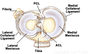
PCL has two inseparable bundles; wider anterolateral bundle (AL) and smaller posteromedial bundle (PM). The AL bundle is tight in flexion or mid flexion and inward rotation of the knee, while PM bundle is tighter in knee extension and deep flexion.
Mechanism of injury
The posterior cruciate ligament can be injured because of various mechanism or excessive movement around the knee joint. It typically requires a powerful force.
- A direct blow to the anterior aspect of the proximal tibia when the knee is flexed. It can easily translate the tibial platform posteriorly.
- Contact or non-contact hyperflexion. The most common mechanism of Posterior cruciate ligament injury among athletes.
- ‘Dashboard injury’, anterior aspect of proximal tibia gets a posterior blow when flexed knee hits the dashboard.
- Hyperflexion injuries.
- Simple missteps
However, posterior cruciate ligament injuries are not common as other ligament injuries. Usually, PCL injuries come along with injuries to other structures in the knee joint such as cartilage, other ligaments, and bone.
Clinical presentation of PCL injury
Acute PCL Injury
Clinical presentation usually depends on the severity of injury and injury to other structures involved. Acute injury presents as a vague, minimal pain with minimal swelling, instability and a full range of motion. Often people even do not realize the acute injury to PCL and athletes keep on practicing their sporting activities. Usually, posterior cruciate ligament injury is combined with other ligaments or menisci or bone that is when the symptoms appear in noticeable extents. Patient presents with marked swelling, increased pain in the anterior and posterior part of the knee, reduced range of motion, difficulty walking with a feeling of weakness or instability as well as stiffness.
Chronic PCL Injury
Often people with a chronic PCL injury do not remember or realize the history or mechanism of injury. Patients only come to the clinic when they are finding difficulty in walking with pain and feeling of instability, marked discomfort in the knee during weight bearing in a semi-flexed position. There can be a retro patellar pain and pain in the medial compartment. However, presentation of pain, swelling, and stiffness depends on the degree of associated injuries to other structures like chondral damage, meniscal injury, avulsion fractures, and collateral ligament injuries.
References:
- Baker CL, Norwood LA, Hughston JC (1984). Acute combined posterior and posterolateral instability of the knee. Am J Sports Med 12:204-208.
- DeFranco MJ, Bach BR (2009). A comprehensive review of partial anterior cruciate ligament tears. J Bone Joint Surg Am 91:198-208.
- Haus, J., Halata, Z. (1990). Innervation of the anterior cruciate ligament. Int Orthop, 14(3), 293-296.
- Hogervorst, T., Brand, R. A. (1998). Mechanoreceptors in joint function. J Bone Joint Surg Am, 80(9), 1365-1378.
- Kennedy, J. C., Alexander, I. J., Hayes, K. C. (1982). Nerve supply of the human knee and its functional importance. Am J Sports Med, 10(6), 329-335.
- Mark L. Purnell, Andrew I. Larson, and William Clancy.Anterior Cruciate Ligament Insertions on the Tibia and Femur and Their Relationships to Critical Bony Landmarks Using High-Resolution Volume-Rendering Computed Tomography. Am J Sports Med November 2008 vol. 36 no. 11 2083-2090
- Morgan PM, LaPrade RF, Wentorf FA et al. (2010). The role of the oblique popliteal ligament and other structures in preventing knee hyperextension. Am J Sports Med 38: 550-7.
- Wind WM, Jr, Bergfeld JA, Parker RD. Evaluation and treatment of posterior cruciate ligament injuries: revisited. The American Journal of Sports Medecine. 2004, 32(7):1765–1775.

Evidence Based Physiotherapy Approach for Frozen Shoulder. Post III
Elbow flexor and extensor muscle weakness in lateral epicondylalgia. Clinical commentary