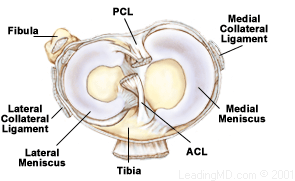
Anterior Cruciate Ligament (ACL)
The knee joint being the joint that plays a key role in weight bearing and mobility, joint integrity or stability is the most important concern. Thus, it is imperative to know and test knee ligaments during any knee joint assessment. Among several ligaments around knee joint, four deserve special attention namely;
- Anterior Cruciate Ligament (ACL)
- Posterior Cruciate Ligament (PCL)
- Medial Collateral Ligament (MCL)
- Lateral Cruciate Ligament (LCL) https://www.youtube.com/watch?v=RTV5Yo3E7VQ
Anterior Cruciate Ligament (ACL)
The anterior cruciate ligament is a band of dense connective tissue which extends superiorly, posteriorly and laterally, twisting on itself as it extends from the tibia to the femur. ACl’s primary functions are to prevent anterior translation of the tibia on the femur, to limit internal rotation of tibia during flexion (open kinematic chain) as it twists around the posterior cruciate ligament. In closed kinematic chain movement during flexion, femoral condyles roll posteriorly, ACL becomes taut, causing condyles to glide anteriorly and limits lateral rotation of the tibia. ACL also checks extension and hyperextension of the knee to a lesser extent. ACL is in relaxed or least stress state between 30 and 60 degrees of knee flexion. https://www.youtube.com/watch?v=_4HJqBtlsEs
Attachments
ACL attaches to anterior intercondylar fossa of the tibia, being blended with the anterior horn of medial meniscus and to the posteromedial corner of the medial aspect of the lateral femoral condyle in the intercondylar notch.
ACL has two bundles; smaller anteromedial bundle (AMB) and larger posterolateral bundle (PLB). The AMB is tight in both flexion and extension, limits anterior translation, and helps stabilize the medial and lateral rotation. The PLB, on the other hand, is tight in extension and medial rotation and limits anterior translation, hyperextension, and rotation.
Nerve Supply
ACL is supplied by the nerve fibers from the posterior articular branches of the tibial nerve. The proximal part of ACL contains mechanoreceptors which provide the afferent arc for signaling knee postural changes. Stimulation of these afferent nerve fibers in ACL can influence motor activity in the muscles around the knee joint; a phenomenon known as Arc Reflex. In the case of ligament injuries, these mechanoreceptors in ACL are affected and thus affecting muscle activations around the knee joint.
Blood supply
ACL gets blood supply by a middle geniculate artery.
Mechanism of injury
ACL can be injured because of various mechanism or excessive movement around the knee joint;
- Anterior blow to tibia resulting in knee hyperextension
- Non-contact hyperextension
- Noncontact deceleration, with tibial medial rotation or femoral lateral rotation on fixed tibia
- Forced tibial medial rotation or femoral lateral rotation on fixed tibia
- Hyperflexion injuries
Rupture of AMB causes anterolateral instability, rotational instability, and increased anterior translation of tibia in flexion whereas rupture of PLB causes an increase in hyperextension, increase in anterior translation, and medial rotation in knee extension in a closed chain.
References:
- DeFranco MJ, Bach BR (2009). A comprehensive review of partial anterior cruciate ligament tears. J Bone Joint Surg Am 91:198-208.
- Haus, J., Halata, Z. (1990). Innervation of the anterior cruciate ligament. Int Orthop, 14(3), 293-296.
- Hogervorst, T., Brand, R. A. (1998). Mechanoreceptors in joint function. J Bone Joint Surg Am, 80(9), 1365-1378.
- Kennedy, J. C., Alexander, I. J., Hayes, K. C. (1982). Nerve supply of the human knee and its functional importance. Am J Sports Med, 10(6), 329-335.
- Konishi, Y., Fukubayashi, T., Takeshita, D. (2002). Possible mechanism of quadriceps femoris weakness in patients with the ruptured anterior cruciate ligament. Med Sci Sports Exerc, 34(9), 1414-1418.
- Mark L. Purnell, Andrew I. Larson, and William Clancy.Anterior Cruciate Ligament Insertions on the Tibia and Femur and Their Relationships to Critical Bony Landmarks Using High-Resolution Volume-Rendering Computed Tomography. Am J Sports Med November 2008 vol. 36 no. 11 2083-2090
- Wu JL, Seon JK, Gadikota HR, et al. (2010). In situ forces in the anteromedial and posterolateral bundles of the anterior cruciate ligament under simulated functional loading conditions. AM J Sports Med. 38:558-563.


One Comment